News
AI Detects Thousands of Hidden Brain Lesions in Multiple Sclerosis
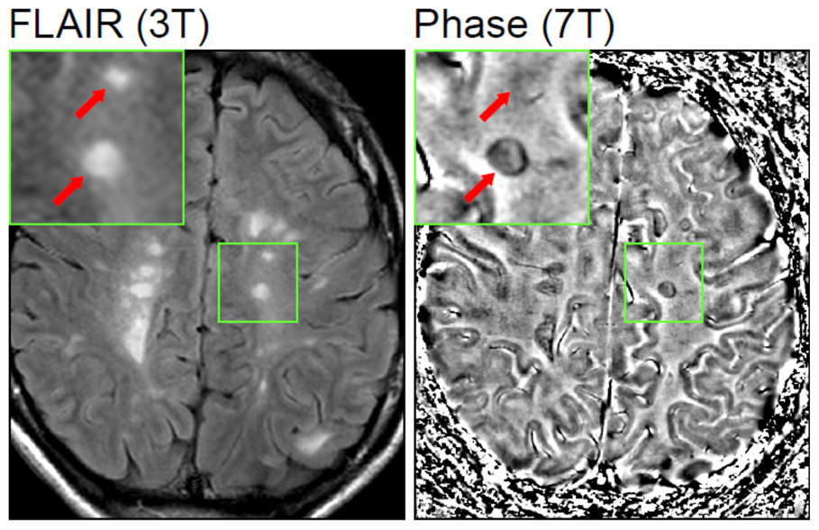
Scientists at the University at Buffalo have developed an AI system that identified more than 11,000 previously invisible lesions in the cerebral cortex of multiple sclerosis patients. The method could change how the disease is diagnosed and monitored worldwide.
Contents
A team at the University at Buffalo has developed an artificial intelligence system that detects lesions in the cerebral cortex of multiple sclerosis patients that were previously invisible on standard MRI scans. The findings were published on July 7, 2026, in the journal Communications Medicine.
The system, called MMCLE (Multimodal Cortical Lesion Enhancement), is a generative AI model that combines several image-processing techniques simultaneously. It uses the FLAIR2 sequence, the T1-to-T2 signal ratio, and a computationally generated double inversion recovery image, which normally requires a separate, time-consuming scan.
How the New Method Works
The key difference from existing tools is that MMCLE analyzes relationships across multiple MRI image contrasts simultaneously, rather than evaluating each scan separately. This allows it to pick up microscopic tissue discrepancies that are missed when individual sequences are assessed the traditional way.
Automatic lesion detection relies on semantic segmentation based on a transformer architecture, the same family of models that underpins large language systems. In this case, the model learns to recognize patterns of abnormal tissue from thousands of previously labeled examples.
The researchers tested the tool on data from the ORATORIO trial, a large international clinical study of primary progressive multiple sclerosis. That database provided the group of 732 participants in whom the system found lesions invisible to standard radiological evaluation.
Why the Cortex Is Hard to Examine
Lesions in the cerebral cortex, unlike those in white matter, are much harder to detect on conventional MRI scans because they have lower contrast against surrounding tissue. Until now, doctors have had to rely mainly on specialized, expensive imaging protocols available at only a few research centers.
The authors emphasize that MMCLE works on conventional, archival MRI scans, meaning data that hospitals and research centers already have on hand. That means the method could be applied retrospectively to analyze thousands of existing databases without requiring patients to undergo new scans.
Detecting previously invisible cortical lesions on conventional, archival MRI scans has serious implications for multiple sclerosis research and clinical care - Dr. Robert Zivadinov, senior author of the study, University at Buffalo
Implications for Diagnosis and Treatment
The number and location of cortical lesions is directly linked to disability progression in multiple sclerosis patients, so detecting them more accurately could help doctors assess treatment effectiveness faster and adjust therapy accordingly. Until now, many patients with real cortical damage may have received results described as stable, despite their disease progressing.
The study was led by Dr. Michael G. Dwyer as first author and Dr. Robert Zivadinov as senior author, in collaboration with researchers from MS Center Amsterdam and Genentech, which sponsored part of the clinical data used in the analysis.
For Polish neurology centers and multiple sclerosis treatment facilities, which together treat more than 45,000 patients nationwide, tools like this could eventually enter clinical practice through integration with existing MRI image-analysis software, without the need to purchase new equipment. However, this requires validation in a broader population and regulatory approval, so commercial deployment remains a distant prospect.
The team plans further work to validate the system in larger, more diverse patient groups, including populations beyond the ORATORIO trial, as well as integration of the tool with existing hospital image-archiving systems.