News
Routine Mammograms with AI Detect Hypertension, Heart Disease and Stroke Risk
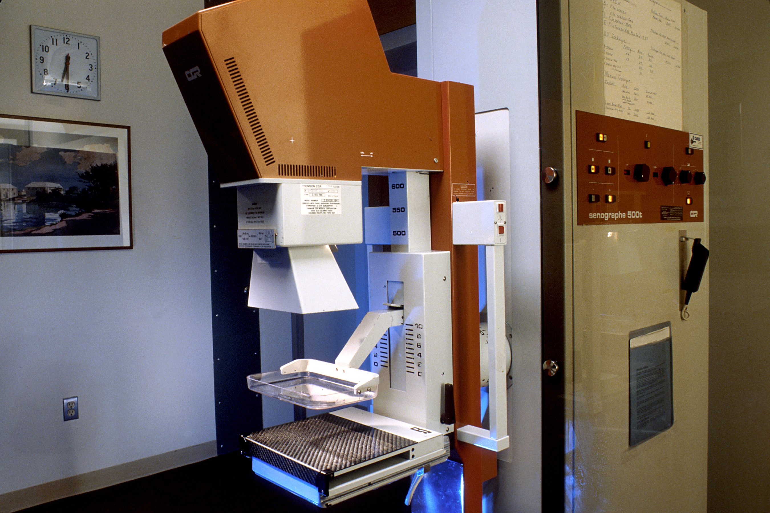
Israeli researchers presented findings at the ESC 2026 congress showing that an algorithm analyzing standard mammography images can identify the three most common cardiovascular diseases in women without additional imaging tests.
Contents
A mammogram, the kind a woman already schedules every year to screen for breast cancer, can also reveal whether she is at risk of hypertension, coronary heart disease, or stroke. That is the conclusion of a study presented on August 27, 2026, at the European Society of Cardiology (ESC) congress in Madrid by a team from Chaim Sheba Medical Center and Tel Aviv University.
The study's author is Dr. Viana Copeland of Chaim Sheba Medical Center, affiliated with Tel Aviv University. Her team trained a deep learning model on nearly one hundred thousand mammograms, teaching it to recognize patterns in breast tissue and blood vessels linked to the three most common cardiovascular diseases in women: hypertension, coronary heart disease, and stroke.
How the Model Works
The starting point was ordinary breast X-rays taken as part of routine cancer screening, with no additional testing involved. The algorithm looked for microscopic calcifications in the walls of the breast arteries, long associated with overall cardiovascular health but typically overlooked by radiologists in daily practice, who focus solely on glandular tissue for signs of tumors.
Performance was measured using the AUROC metric, which shows how well a model distinguishes sick patients from healthy ones on a scale from 0.5 (chance) to 1.0 (perfect discrimination). For stroke, the score was 0.86, for hypertension 0.79, and for coronary heart disease 0.78, levels comparable to many standard screening tools currently used in cardiology.
Women Overlooked in Diagnostics
Heart disease remains the leading cause of death among women worldwide, yet it is systematically underdiagnosed and undertreated in women compared with men. Symptoms can be atypical, and classic cardiovascular risk scales were developed mainly using data from men. Mammography, meanwhile, reaches millions of women every year as a routine preventive test, so adding cardiovascular risk analysis to it requires no extra visit or radiation exposure.
Since mammography is already widely used, analyzing the same images for cardiovascular health information could potentially offer a scalable approach without requiring additional imaging - Dr. Viana Copeland, Chaim Sheba Medical Center
Earlier Confirmation in a Large Sample
The new work builds on ground prepared by an earlier study from a team at Emory University led by Dr. Hari Trivedi, published in March 2026 in the European Heart Journal. It covered 123,762 women aged 40 to 79 from two large U.S. healthcare systems, none of whom had a previously diagnosed heart condition at the time of the study.
That model measured the amount of calcification in the breast arteries visible on a mammogram and translated it into future risk of serious cardiovascular events. Women with mild calcification had roughly a 30 percent higher risk of serious heart disease than women with none, moderate calcification raised the risk by more than 70 percent, and severe calcification was associated with a two- to threefold higher risk. The association held even among women under 50, usually considered a low-risk group, and remained significant after accounting for factors such as diabetes and smoking.
What Comes Next for Deployment
Neither tool is yet approved for routine clinical use. Both studies are observational and require validation in prospective trials before they can enter radiologists' practice. The key question is whether an algorithm's output will actually change a doctor's course of action: will a patient with a high-risk score be referred for further cardiac evaluation, or will it simply add one more number to the report that nobody knows how to interpret.
For healthcare systems, the appeal of this approach lies in cost: analyzing images that are already being taken requires no new equipment or extra time from the patient, just software layered onto data already collected. It's a model similar to other AI applications in imaging diagnostics, where an algorithm adds a second layer of information to a scan performed for an entirely different reason.
In Poland, mammography under the Populacyjny Program Wczesnego Wykrywania Raka Piersi (the national population-based breast cancer early detection program) covers women aged 45-74 and is performed every two years, meaning a potentially large population on which a similar tool could be used, should it be validated and approved for use beyond the United States and Israel, where the two cited studies originate.