News
Berlin Scientists Train AI to Analyze Microscopic Lung Mucus Images
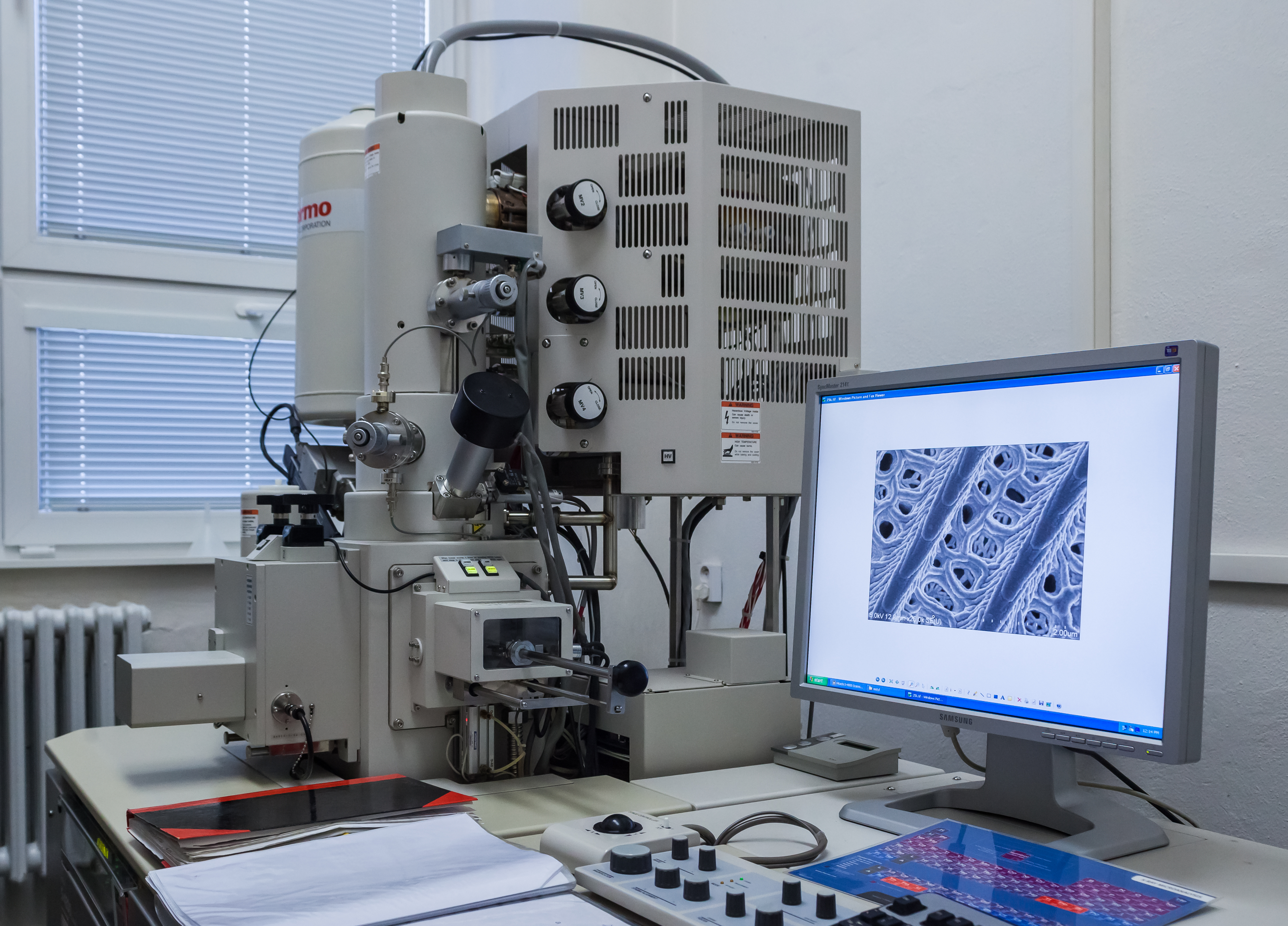
A team from Charité and Freie Universität Berlin built an AI system that assesses the structure of mucus from the airways and intestines in electron microscope images 40-120 times faster than a human expert, opening the door to faster diagnosis of cystic fibrosis and COPD.
Analyzing a single electron microscope image, in which pores and fibers in the mucus mesh must be measured by hand, takes an experienced researcher 10 to 30 minutes. A team of scientists in Berlin has shown that a machine learning system can do the same job in about 15 seconds, describing the results in the journal American Journal of Physiology-Lung Cellular and Molecular Physiology.
How the system works
The tool combines a classic imaging technique, scanning electron microscopy (SEM), with the Trainable Weka Segmentation algorithm running on Fiji, a platform popular among biologists. A multi-layer perceptron neural network classifies every pixel of the image into one of four categories: dark pores, light pores, dark fibers, and light fibers.
Based on this, the system generates probability maps, which are then converted into binary images. From these simplified maps, the algorithm calculates specific structural parameters of the mucus network: pore size, fiber length, and the degree of branching in the mesh.
Why mucus structure matters
Mucus in the airways and intestines acts as a filter and protective barrier, and the density of its pore network determines how easily bacteria, viruses, or drugs can pass through it. In diseases such as cystic fibrosis, chronic obstructive pulmonary disease (COPD), or severe asthma with mucus overproduction, this structure changes, making the mucus harder to clear from the body and encouraging infections.
Until now, such measurements were done by hand, which limited the number of samples that could be analyzed in a single study and introduced variation stemming from individual researchers' subjective judgment. Automating the process is meant to make it possible to analyze much larger datasets in a reasonable amount of time.
Limitations of the study
The paper's authors, among them Yannic Kerkhoff and Matthias Ochs listed as corresponding authors, caution that the tool has so far only been tested on samples from healthy donors. It has not yet been validated on material from cystic fibrosis patients, the group for which a clinical application would be most valuable.
That means that before the method can move into diagnostic practice or research on new mucus-thinning drugs, the analysis will need to be repeated on samples from patients and the results compared against existing methods for assessing the condition of patients' airways.
What comes next
The system runs locally as a plugin for Fiji, a free platform for analyzing microscope images used widely in biology labs around the world, which lowers the barrier to entry for other teams that want to use it without building their own computing infrastructure.
The work reflects a pattern that has repeated across biomedical science in recent months: machine learning models here are not replacing doctors or researchers, but taking over tedious, repetitive measurement work, freeing specialists to focus on interpreting results and designing further experiments.